navicular bone fracture symptoms
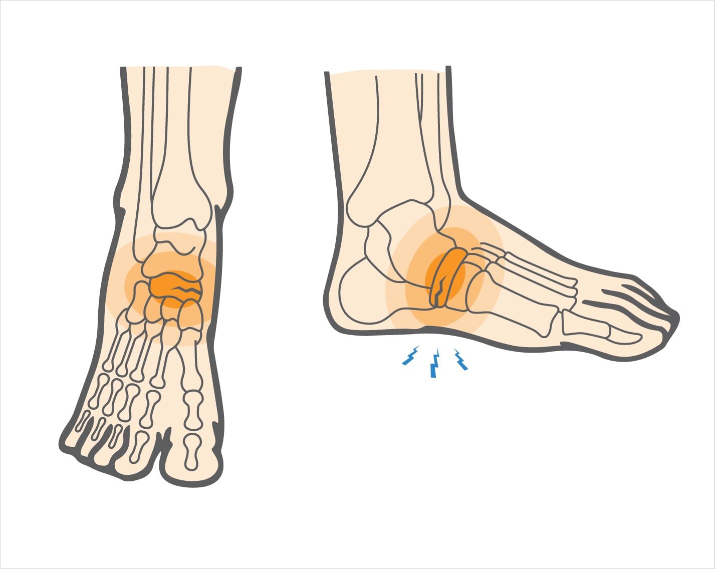 Navicular Stress Fracture | Upswing Health
OF THE WORKING SCIENCE Silence MENU Follow Us Molecular Stress Fracture: A High Impact Risk for Young athletes Chris Mallac investigates the causes, diagnosis and management of navigable stress fractures in athletes. Team GB Triple Jumper Benjamin Williams during a training session in Tunstall Park, following the outbreak of coronavirus disease (COVID-19), May 2020. REUTERS/Carl RecineFirst described by Towne and colleagues in 1970(1), navicular bone stress fractures are rare in the general population. However, male athletes in the mid-20s who participate in sports such as sprinting, average running distance, hug and basketball are more at risk(2,3). These particular sports subject the player to high foot and middle foot forces when jumping, landing, running and cutting (4,5). Elite-level athletes comprise 59% of the navigable stress fractures reported in athletes with average foot pain. These injuries tend to occur after peaks in training (higher mileage) and racing loads(6). Anatomy and biomechanics The navicular bone is wet and placed between the three quaneiforms of the middle foot and the head of the stem on the back foot. It is a concave-shaped bone with the concave surface articulating with the talus (see figure 1). The posterior tibialis tendon inserts into the medial tuberosity of its medial surface. The other significant anatomical characteristic is the plant peak, to which the calcaneonavicular plant, or spring, ligament aggregates(7). Because it is connected to the head of the proximally talus and the three quaneiforms in a distual way, the navicular bone forms a pivotal link between the middle foot and the rear foot for the transmission of force and thrust(8). Being located in the foot's longitudinal arch, it also plays an integral role in maintaining the integrity of the arch(4). Its position between the talus and the 1st and 2nd cuneiform means that the nivico supports additional cutting and compression forces during operation and landing. In particular, the central third of the bone holds focused lateral cutting forces, which can predispose it to a stress fracture (4.9). Subsequent thybialis muscles, which are inserted into the nivico, can exacerbate the vulnerability to stress in the region through their traction forces in the bone. The medial nerve plants internalizes the thalavicular joint. Therefore, pain can refer to the foot ball imitating Morton's neuroma when the navico is the source of pain. Figure 1: Fracture of anatomy and nautical stressIn addition, the supply of blood to the nauticals enters the non-artic surfaces of the bone, and is branched in a medial and lateral direction without a large amount of blood supply to the central third. This circulation distribution creates an area of avascular basin in the bone, especially in individuals with genetic predisposition to this morphology (see figure 2)(10). If present, this relative avascular area can lead to delayed healing in case of fracture, and higher rates of non-union(9). Figure 2: Area of avascularity (adaptada de Khan et al. 1994)3 The following are risk factors for developing a vigicular stress fracture(11):Diagnosis Athletes with navigable stress fracture usually report one or more of the following: Figure 3: The N point in the dorsal aspect of the distal thalavicular joint. Diagnostic Confirmation A simple film X-ray is usually ineffective to detect a navicular stress fracture (8.14); most fractures are incomplete, and it takes 10 days to three weeks for bone resorption to be shown in the X-ray if there is a complete gap (3.15). However, the X-ray image may exclude other possible causes of pain, such as a stem neck that stimulates or capsular avulsion. A triphasic bone scan is highly sensitive and has a high positive predictive value for stress fractures in the navicle, showing absorption in all phases(13). However, they are not specific and lack the anatomical resolution to see the size and location of the stress fracture. Magnetic resonance (MRI) is the imaging method to see early changes in the bone (see figure 4). It can detect both bone edema and stress reactions, which occur before a stress fracture. As such, an MRI can take stress reactions before progressing to stress fractures in excess(11). A CT scan can show the exact location and size of a navigable fracture. It is sensitive to partial fractures, which are often coursed from the proximal central-teral pertigo of the bone to its distal planting pole (3). The findings of the CT scan dictate the classification of a navigable stress fracture. However, the poor position and the thick TC slices (more than 2 mm) could be lost a stress fracture(16). The most widely used classification system is Saxena et al. (2000)(17). The three types of stress fracture are: Other classifications based on changes observed in the bone include avascular, cystic or sclerotic subtypes. Figure 4: MRI showing bone edema in the navicle** (adapted and used with permission of )ManagementNormal stress fractures usually progress along a three-stage continuum. The first is a stress reaction, typified by a short duration of pain (less than two weeks), which can decrease with rest. However, patients will have a "N" tender, and while computed tomography is normal, an MRI will show bone edema. Below is the stress fracture phase, where there is constant average foot pain exacerbated by activity. The fracture is evident both in a CT and in a MRI test. Later, the patient can develop a complicated stress fracture, with long-standing pain, even after a non-weight bearing period. The management protocol of these phases is as follows: The treatment attachments at this stage include: Table 1: Management guides for a stress fracture Weeks 0 – 6Non-weight bearing in fiberglass or boot casting. At the end of week 6, perform a CT scan to measure healing (if justified) and evaluate point N. If it is painful, keep the bearing without weight for another two weeks. Weeks 7 – 8Optional full weight-bearing in a CAM boot or total weight-bearing without boot if 'N' spot is non-tender. Weeks 8 – 12Start running on the grass in alternative days. Start with five minutes and increase in five minutes each week. Once the load is executed is up to ten continuous minutes, the progress to the tracks for distances of about 60-80 meters, with a build-up of speed and volume for a few weeks. Weeks 13-16Start light training with a gradual accumulation of load and intensity. Weeks 16+ Return to competition. Although studies are limited, the general findings of the conservative and operational management of vicular stress fractures show that in cases of conventional management, the time to return to sport is about 22 to 26 weeks. While surgically treated tend to return to competition in 15-18 weeks(2,14). ReferencesAbout Chris MallacRelatedin , While practice can make perfect, too much preparation can damage the bones in growth. Chris Mallac explains how launch mechanics and skeletal immaturity contribute to the small league elbow. 2019 South Williamsport, PA, Egan Prather launcher (24) launches a third entry against the Caribbean Region during the World Small League Series. Credit: Evan Habeeb-USA TODAY... in, Sesamoide's bones are some of the smallest in the body. However, they may pose a major problem when they are injured. Trevor Langford discusses anatomy, biomechanics and clinical relevance of a stress fracture of the sesameid bones and reviews management options. Sports such as running, dancing and gymnastics often require a forcible dursiflexion while in a weighing... in , , athletes with persistent and indiagnosed shoulder pain can suffer rare but painful quadrilateral spatial syndrome. Chris Mallac unravels this diagnostic complex and offers practical treatment solutions. The Quadrilateral Space Syndrome (QSS), first described in 1983, is an unusual and unusual shoulder pathology that is seen mainly in male volleyballists, swimmers and baseball launchers(1,2). Athletes with QSS... in , , In the second part of this two-part article, Andrew Hamilton examines the best imaging patterns for hip avulsion injuries and explores the most up-to-date guidelines for injury management. As discussed in the first part of this article, the presentation and location of a hip avulsion lesion may vary a lot. This is due to... in, , In the first of a series of two parts, Chris Mallac explains the functional anatomy of the major pectoralis and their tendon, the situations that put the tendon at risk of injury, and the signs and symptoms of a broken tendon. The first reported case of pectoralis mayor (PM) tendon rupture occurred in Paris in 1822. Above... in, Tracy Ward considers the biomechanical and physical aspects of the paddle and its contribution to the lesions of the upper extremity and explores injury prevention strategies, including specific strength and conditioning. Rowing is a repetitive and weight-supported sport where high training volumes are similar to cycling, kayaking and swimming(1). Despite being non-contact and low-impact, runners are still subject to... Latest articles "ReLEVANT and CONCISE Information" "ReLEVANT and CONCISE Information" Robert Barton, D.C. Peak Performance Spine & Sports Medicine Quick Links Most Popular The Sports Injury Bulletin brings together a global panel of experts, including physical therapists, doctors, researchers and sports scientists. Together we deliver everything you need to help your customers avoid – or recover as quickly as possible – injuries. We clear the scientific jargon and deliver easy-to-follow training exercises, nutritional advice, psychological strategies and recovery programs and exercises in clear English.© 2021 Newsletter of Sports InjuriesPart of Green Star Media Ltd. Company number: 3008779
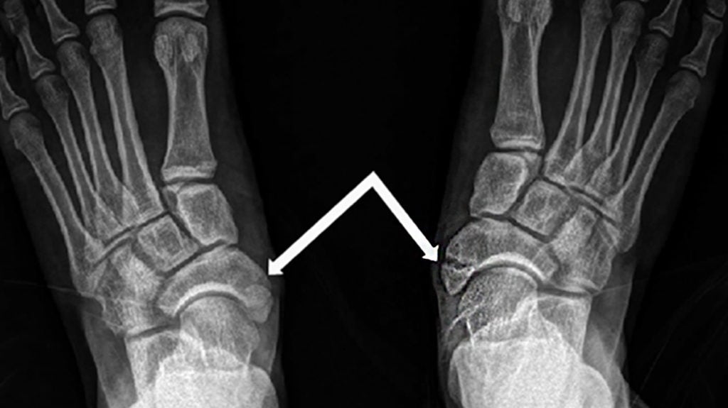
Navicular Fracture in Foot and Wrist

Navicular stress fracture: a high-impact risk for young athletes

Navicular stress fractures & the phases of bone remodelling — Rayner & Smale

Navicular Fracture | Fracture symptoms, Sports massage, Rehab

Key Insights For Treating Navicular Stress Fractures | Podiatry Today
Stress Fractures: Causes, Symptoms, Tests & Treatment

Navicular stress fracture: a high-impact risk for young athletes
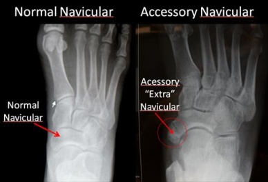
Accessory Navicular Bone - Physiopedia

How can you quickly recover from a navicular stress fracture? | Dr. David Geier - Sports Medicine Simplified

Tarsal Navicular Stress Fractures - American Family Physician

Tarsal Navicular Fractures - Foot & Ankle - Orthobullets

Navicular fracture | Radiology Reference Article | Radiopaedia.org

Navicular Bone - an overview | ScienceDirect Topics

Navicular stress fractures & the phases of bone remodelling — Rayner & Smale

Navicular stress fracture: Signs, symptoms and treatment options | Dr. David Geier - Sports Medicine Simplified

Navicular stress fracture: Signs, symptoms and treatment options - YouTube

Tarsal Navicular Stress Fractures - American Family Physician

Navicular stress fracture: a high-impact risk for young athletes
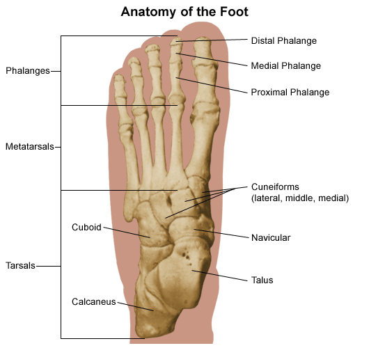
Sports and Fractures | Johns Hopkins Medicine
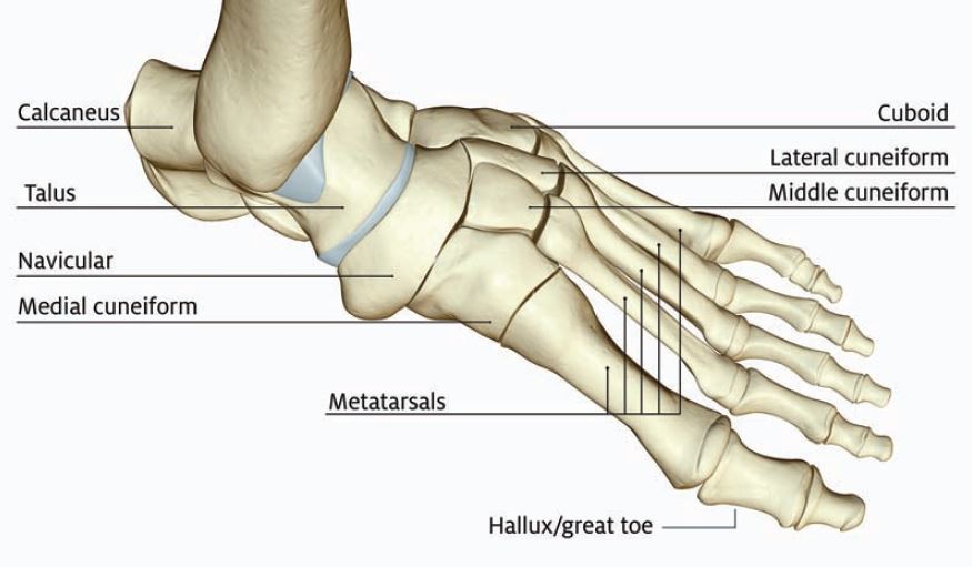
Managing Foot Fractures in Urgent Care | Journal of Urgent Care Medicine
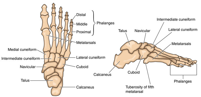
Navicular Stress Fracture - Symptoms, Causes, Treatment & Rehabilitation

Accessory Navicular Syndrome – Caring Medical Florida
Navicular Fracture - Western New York Urology Associates, LLC

How can you quickly recover from a navicular stress fracture? - YouTube
Tarsal Navicular Fracture in a Parkour Practitioner, a Rare Injury - Case Report and Literature Review
/talus-fractures-2549436_final-3b5774c8102f4aa58615e0df5e2af0f7.png)
Talus Fracture of the Ankle Overview
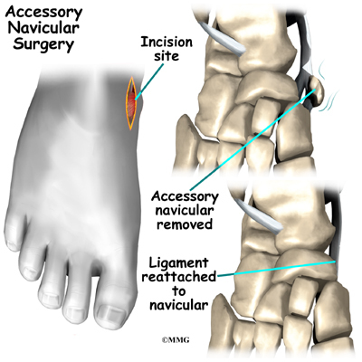
Accessory Navicular Problems | Orthogate
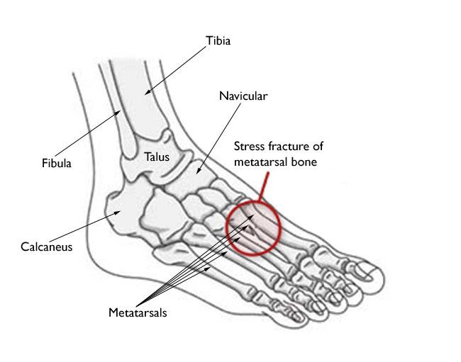
Stress Fractures of the Foot and Ankle - OrthoInfo - AAOS

Accessory Navicular Problems | 2020 OrthoNorCal, Los Gatos, Capitola, Morgan Hill, Watsonville, CA
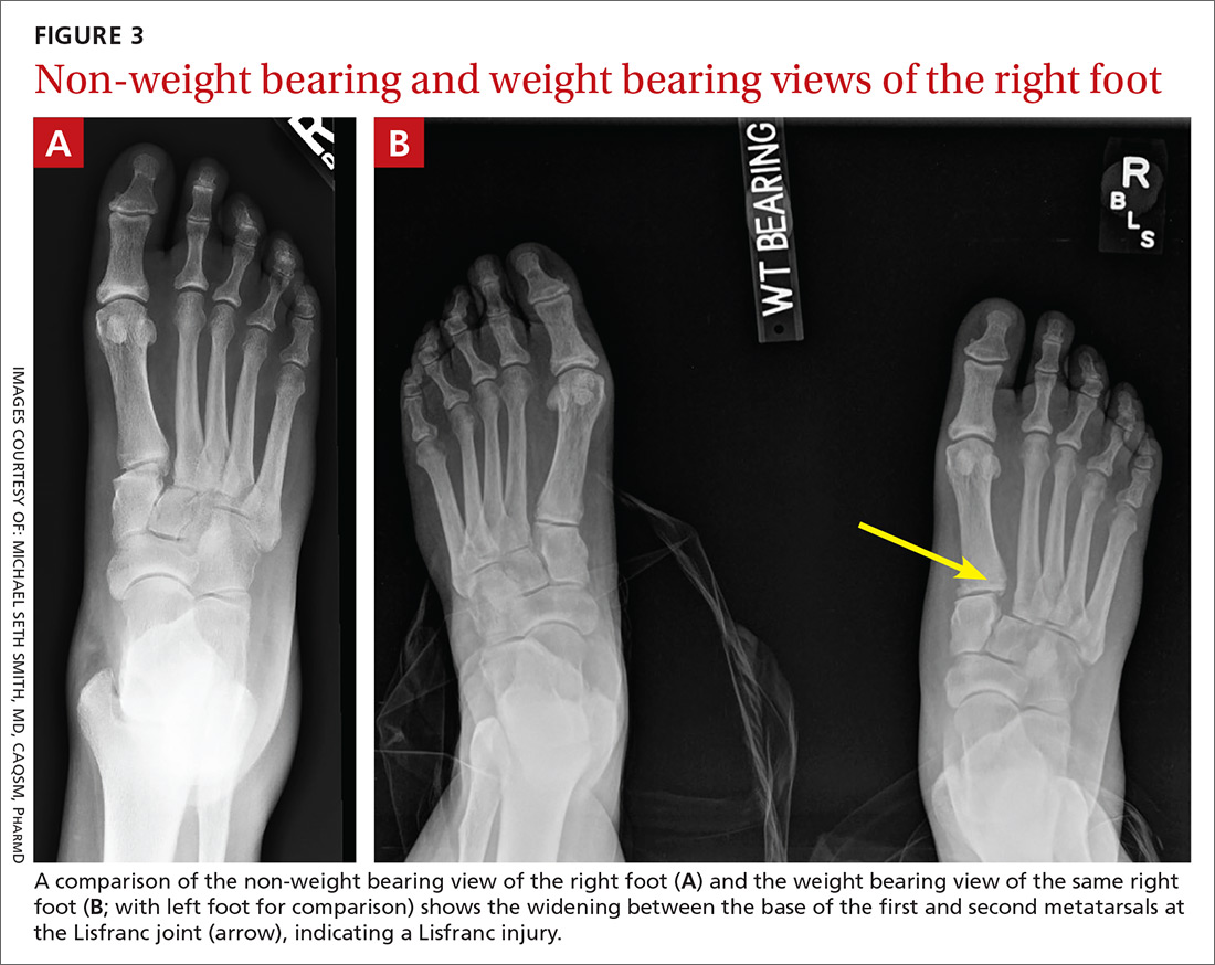
Adult foot fractures: A guide | Clinician Reviews
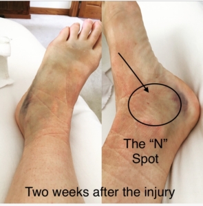
Nursing a Navicular and Talar Head Fracture | GOOD MOXIE

Tarsal Fracture|Signs|Symptoms|Treatment-Cast, NSAIDs, Surgery, Rehab, Exercises
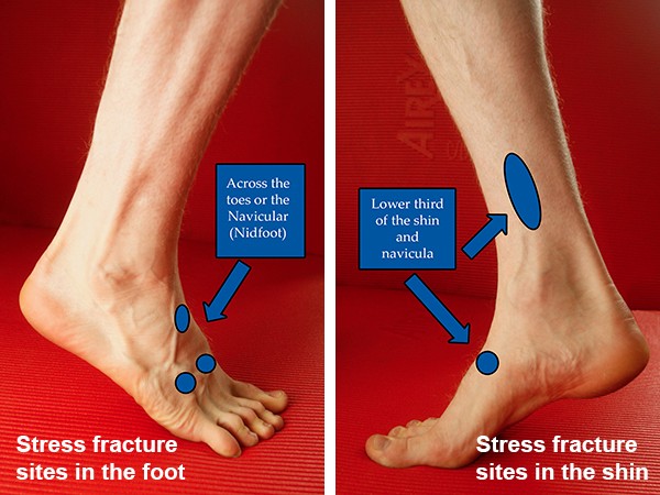
Stress Fractures – Diagnosis And Recovery | realbuzz.com

A 51-year-old female patient with an avulsion fracture of the navicular... | Download Scientific Diagram
Tarsal Navicular Fracture in a Parkour Practitioner, a Rare Injury - Case Report and Literature Review

Navicular Fractures - Everything You Need To Know - Dr. Nabil Ebraheim - YouTube
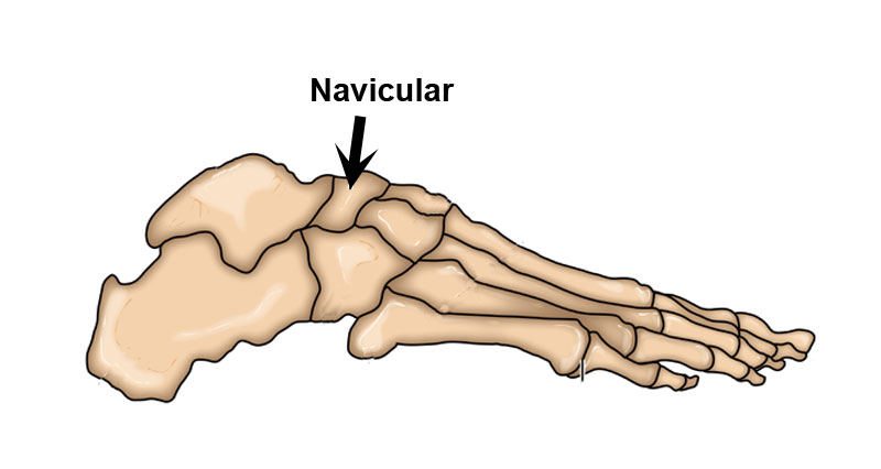
Navicular Stress Fracture - Symptoms, Causes, Treatment & Rehabilitation
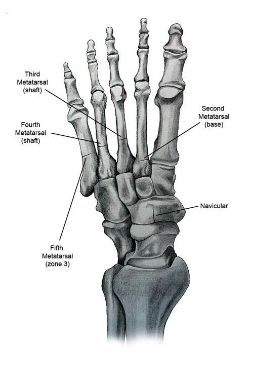
Foot & Ankle Stress Fractures: Causes, Symptoms, Treatments

What are the symptoms of a hairline fracture? - New Mexico Orthopaedic Associates, P.C
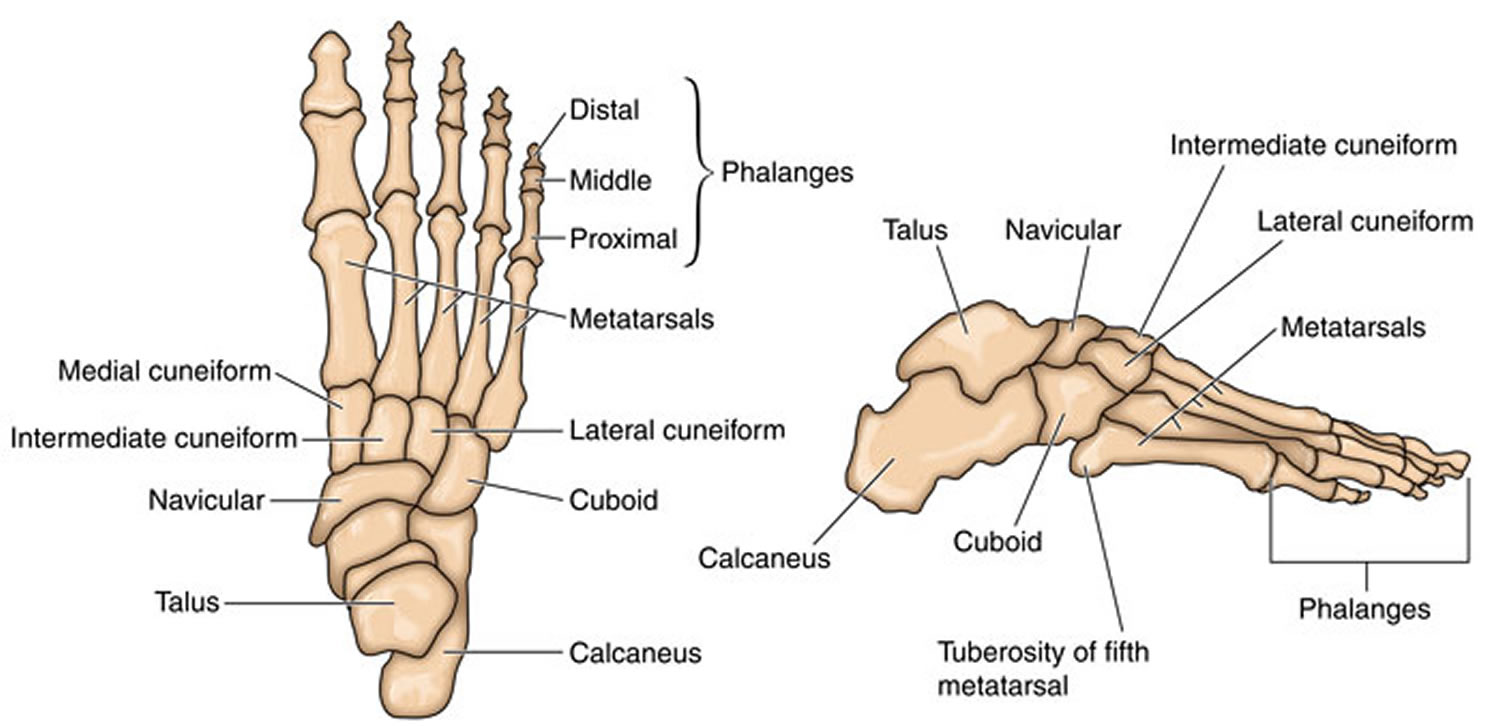
Navicular fracture causes, symptoms, diagnosis, treatment & prognosis
Posting Komentar untuk "navicular bone fracture symptoms"